The frenetic pace of change in the field of medical imaging has seen a raft of new developments take centre stage to improve patient diagnosis and treatment. Thanks to advancements in digital technology and increased investment into medical imaging research, hospitals around the world are benefiting from techniques that allow for enhanced resolution, increased efficiency and improved safety for patients.
From ultrasound and computed tomography to new medical resonance imaging modalities, the past few decades have seen the tools of radiologists evolve to provide more accurate diagnoses and improve patient care. Already, 2018 has seen a number of ground-breaking developments in medical imaging prove revolutionary to both radiologists and patients – the month of November alone bringing a few ‘firsts’ in this critical arm of healthcare:
1. ADVANCED IMAGING TECHNOLOGY MEASURES MAGNETITE LEVELS IN THE BRAIN
In 1992, researchers discovered the presence of magnetite – permanently magnetic form of iron oxide—in human brain tissue. While iron is commonly found in the human body, the presence of magnetite could be cause for concern, with numerous studies linking heightened levels of magnetite in the brain with Alzheimer’s disease and other neurodegenerative diseases. Previously, the identification of magnetite particles was only possible in post-mortem brains – until now.
In November 2018, researchers at Martinos Center for Biomedical Imaging at Massachusetts General Hospital (MGH) harnessed magnetoencephalography (MEG) — an advanced imaging technology that measures brain activity by detecting the weak magnetic fields produced by the brain’s normal electrical currents – to detect and map the presence of magnetite in a living brain. With further research, it’s thought that radiographers could eventually detect the development of Alzheimer’s in a patient by measuring the level of magnetite particles in the brain. According to David Cohen, PhD of the Martinos Center, it poses the question as to whether treatments that influence magnetite could alter disease progression.
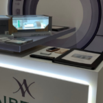
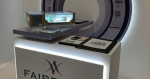
2.THE FIRST FULL-BODY MEDICAL SCANNER
Following a decade of development, the first full-body scanner has finally produced its first 3D image of the entire human body in one instant scan. Developed by the University of California Davis scientists Simon Cherry and Ramsey Badawi, ‘Explorer’ is a combined positron emission tomography (PET) and x-ray computed tomography (CT) scanner. Already, the results are being described as revolutionary in assisting both clinical research and patient care.
Having conceptualised their full-body scanner over 13 years ago following a $1.15 million grant from the National Cancer Institute, Badawi and Cherry were reportedly dumbfounded by the results of their creation. According to Simon Cherry, the potential of Explorer lies in its ability to produce higher-quality diagnostic PET scans than have ever been possible. What’s more, it also completes scans 40 times faster than current PET scanners and can scan with a radiation dose 40 times less than PET scans. With the opportunity to enhance safety and improve resolution, accuracy and speed all at once, the development of Explorer is nothing short of a breakthrough,
3. NEW MRI METHOD PROVES REVOLUTIONARY IN DETECTING LONG-TERM BRAIN DAMAGE
In the UK, a study of over 200 babies at seven hospitals found a 15-minute magnetic resonance (MR) spectroscopy scan was capable of predicting brain damage with 98 percent accuracy. At present, any baby suspected of having brain damage will undergo an MRI scan shortly after birth, allowing doctors to identify any anomalies in the brain through black and white images. The information gained from the scan enables doctors to inform parents on the potential long-term consequences of the damage. As valuable as the MRI scan has been in detecting signs of damage, this technique only offers 60-85% accuracy, and doctors can only confirm if a child has lasting brain damage when they reach the age of two by assessing their development progress.
Led by Imperial College London, the new study used MR spectroscopy to determine the health of brain cells in the thalamus by testing for N-acetylaspartate, a compound which can be found in high levels in healthy brain cells.
Dr Sudhin Thayyil, leading author of the study and Director of the Centre for Perinatal Neuroscience in Imperial’s Department of Medicine said: “At the moment parents have an incredibly anxious two-year wait before they can be reliably informed if their child has any long-lasting brain damage. But our trial – the largest of its kind – suggests this additional test, which will require just 15 minutes extra in an MRI scan, could give parents an answer when their child is just a couple of weeks old. This will help them plan for the future, and get the care and resources in place to support their child’s long term development.”