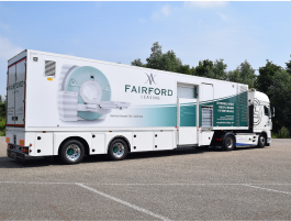
Medical Resonance Imaging (MRI) machines play a critical role in modern healthcare – so much so, in fact, that most of us take for granted the capabilities they provide to hospitals and healthcare facilities across the world. With its ability to map the internal organs and functioning of the human body without the use of X-rays, MRI is well accepted as one of the biggest medical breakthroughs of all time.
As the name suggests, MRI uses a powerful magnetic field and radio waves to produce detailed images of anatomy and pathology inside the human body, making it an invaluable tool in the diagnoses of illness and injury. However, unless you have had experience of an MRI machine or work in the field of radiology, you may not appreciate the long list of use cases that a scanner of this variety has in diagnosing anatomical and pathological anomalies within the body:
IDENTIFYING ANOMALIES IN THE BRAIN AND SPINAL CORD
From degenerative diseases such as Multiple Scleorisis to fluid build-up in the brain, tumours and Pituitary gland disorders, MRI scans enable physicians to detect the presence of structural abnormalities or other conditions in the brain and spinal cord.
Functional MRI of the brain (FMRI) is used to pinpoint the exact location of the brain where a function such as speech happens. When you undergo an MRI imaging of the brain, a radiologist will ask you to perform a particular task while the scan is taking place. In doing so, physicians can determine the exact location of the functional centre in the brain and are well-equipped to offer personalised treatment plans.
INJURIES TO MUSCLES, BONES, LIGAMENTS OR CARTILAGE
If you are experiencing pain, weakness, tingling or swelling around a muscle or joint, an MRI scan will allow your doctor to get to the root cause of the problem by capturing an accurate image of your bones, cartilage, tendons, ligaments, muscles, and even some blood vessels. MRI test results can reveal a number of problems causing the pain such as damaged cartilage, torn tendons or ligaments, bone fractures, osteoarthritis or tumours.
ASSESSING POTENTIAL HEART CONDITIONS
If a patient complains of heart problems, a cardiac MRI scan can be used to view the structure of the heart and assess how well it’s pumping blood. A cardiac MRI could be used to assess age-related wear and tear of the heart valves or determine the damage to the heart following cardiac arrest.
Unlike an X-Ray, an MRI scan does not use radiation so is therefore dubbed the safer and more accurate method of mapping the heart. They are also commonly used to assess the blood supply to the heart, which can enable doctors to uncover conditions such as coronary heart disease or the underlying reasons for reduced blood flow to the heart.
DETERMINING THE CAUSE OF DIGESTIVE ISSUES
Digestive disorders affect billions of people with the severity of conditions ranging from minor to potentially life-threatening. As such, MRI scans are regularly used by healthcare providers to better understand what is causing a patient to suffer digestive problems, whether it’s a food intolerance, a chronic issue such as Irritable Bowel Syndrome or something more serious such as cancer or a hiatal hernia.
When patients experience symptoms such as bleeding, bloating, pain, nausea or vomiting, doctors will commonly call on radiologists to undertake a non-invasive MRI scan in order to get an early diagnosis and put together the most appropriate plan for treatment. Beyond diagnosing diseases, this medical imaging modality can further be used to monitor treatment for a variety of digestive conditions.


